Devina Kothari
Design Innovations
Client: Sanofi, U.S.A
Concept Name: Being 'Sweet' isn't 'Painful'
Description:
“Being sweet, isn’t painful” is a concept that disassociates the social taboo associated with diabetes. It integrates the insulin delivery system with the patient’s lifestyle by amalgamating the existing technology advancement with fashion.
The product is characterized by a closed loop delivery of insulin, achieved by two inter-connected units, The Monitoring Unit and The Diffusing Unit.
The Monitoring Unit:
Comprising of the following:

Outer Case

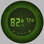
Customised detailed view of the LED display of the Monitoring Unit.


Stainless Steel
Probe
Physical Connector
LED Display
(Customised detailed view)
Microprocessor
1.
3.
2.
4.
Outer Case
Micro-Needle Array
Physical Connector
Cavity for Insulin Reservoir
Microprocessor


This unit is attached to the patient with the help of a 316L stainless steel Probe, which functions as a biosensor and continuously detects and monitors the blood glucose level and the pulse rate, with the help of an embedded microprocessor. The microprocessor receives inputs from the probe and transcodes it into relevant inputs for The Diffusing Unit, thereby continuously adjusting the diffusing units of insulin (basal and intermittent bolus) as per the need of the body.
The micro-processor is also connected to the back-lit LED which displays:
-
The current glucometer reading along with %change
-
Current battery reserve of the micro-processor
-
Insulin content in the reservoir
-
Pulse rate
The Monitoring Unit is physically connected to the Diffusing unit. In the absence of the Monitoring Unit, the Diffusing Unit still functions in isolation as a stand-alone device, with manual feed.
Visually the monitoring unit is differentiated from the diffusing unit by a back lit LED display and a thinner cross section. This is very useful for visually impaired patients.
The Diffusing Unit:
Comprising of the following:
The Diffusing unit is adhered to the patient’s body through an array of needles. This silicone micro-needle array is used to diffuse insulin. An array of forty eight 400micro-mm needles arranged in eight concentric circles aid in continuous transdermal diffusion of insulin, governed by inputs from it’s microprocessor. The micro needle array provides a pain-free penetration into the patient’s body. It eliminates the pain associated with multiple injections of insulin and provides the patient a pain-free environment for his personal development. The microprocessor of the Diffusing Unit is connected to the microprocessor of the Monitoring Unit, through physical connection. The microprocessor of the Diffusing Unit controls the non-return valve and aids in dispensing precise amount of insulin from the reservoir through the needles into the patient’s body. The base of the reservoir is selectively permeable and allows insulin transmission through absorption. The absorbed insulin is further channelized through a non-return valve that controls the diffusion through array of needles.
The cavity of the diffusing unit hosts the Insulin Reservoir. This reservoir stores large volumes of insulin, up to 100 units (1ml), 200 units (2ml) etc. One may insert the globule into the reservoir as per their personal requirement. For example, a person consuming 50 units of insulin per day would insert a 2ml globule into the reservoir to remain ‘tension-free’ for four days; and then dispose off the system. If an individual is heading towards a trek, or a long journey, secondary Reservoir may be attached to the monitoring unit, which may extend the ‘tension-free’ days without replenishment.
Sites of Application:
The device can be locally administered onto the following sites on the human body:
-
Abdomen (except for the 100mm diameter around the navel).
-
Top and outer thighs (avoiding the boney area around the knees).
-
Outer and upper arms (outer back area of the upper arm, around the fatty tissue).
Size:
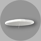
The Diffusing and Monitoring Units are of equal outer diameter, being 50mm. The monitoring unit is thinner to the diffusing unit. The former being of 5mm and the latter being of 10mm. This aids the visual impaired patients to differentiate between the two units easily.
The connectors are 2mm thick, and keep the units within the radius of a maximum of 75mm from each other.
Form:
Circular form renders the device a neutral appeal with respect to gender and age. Absence of sharp edges helps to keep the garments safe from tear or entangle and avoid unsolicited nudging. The device is thinner than an adult human finger, enabling it to be concealing under the garments without visible outer contour.
Colour:
The device is integrated as a lifestyle accessory. The backlit LED display aids in changing colours as per the desire of the patient rendering a style statement in case of visibility onto the skin. It can be clubbed with the accessories adorned by the patient. The outer casing has a glossy finish and the Silicone rubber cased connector matches with the patient’s skin tone.
Materials and Coatings :
-
Silicone based multi-layer liquid repelling nano-coating on the electronics to render the system waterproof. This aids in normal course of daily routine, like bathing, swimming, exercising, etc. with both the units continuously attached to the patient’s body.
-
Areas that come in contact with the patient’s body, are treated with inert materials, to avoid skin irritation or reaction with sweat and other secretions.
-
316L Stainless Steel (Surgical grade) Probe to be inserted into the human body.
-
Use of Platinum cured Silicone for outer casing.
-
Primary and Secondary Diffuers are attached to the Monitoring Unit, for continuous transmission of Insulin for extended priods without replenishments.
Local administration sites for Insulin diffusion.

Monitoring and Diffusing Units conncted and adhered on to the patient's body.

Nano-Coating of the Silicone Rubber on to the electronic circuit.


Top and Bottom views of the two Units. The Monitoring Unit is thinner than the Diffusing Unit.
Insulin Pen used to replenish the Diffusing Unit, in case of unavailability of Insulin Globules.
The Monitoring Unit is once paired with five devices for continuous monitoring and future auto-configuration.
2 Enucleation Kit
Probe at 45 degrees from normal for easy insertion into the body.

Advantages:
-
The system is waterproof due to the silicone rubber coating on the electronics.
-
Pre-calibrated Insulin pen can be used to replenish the reservoir in absence of insulin globules. These insulin pens may be paired to the Monitoring Unit of the patient to avoid manual calibration every time. Once paired with the Monitoring Unit of a particular patient, it can be used to pair successive Monitoring Devices of the same patient.
-
The Monitoring Unit of the patient can be paired with the Endocrinologist’s system, to further increase the accuracy of monitoring and delivery of insulin. It can be further paired with three other devices, to which regular alarms or feedback regarding any unusual fluctuation in the patient’s blood glucose level or pulse rate may be sent.
-
Due to the disposability of the two units, pairing of devices is required frequently. Successive monitoring units are paired with any of the previously paired device for auto-configuration.
-
This system is highly useful to infants, children, immobile, unconscious, handicapped, and dependent patients or those under intensive care; as it monitors, diffuses and alarms simultaneously without frequent human intervention. The system of devices is self administered with minimal skills or training and across a broad segment of patients.
-
Pre-charged battery attached to the microprocessor of the monitoring unit renders functionality for extended periods increasing usability in regions with limited supply of electricity. The battery can be also continuously re-charged by the volts generated through the continuous ion (sodium and potassium) exchange inside the human blood stream.
Pairing of Devices

5 steps to a pain-free lifetstyle:


1
Physically connect the Diffusing Unit to the Monitoring Unit.
3
5
Insert the requisite volume of insulin
into the reservoir,
using a blister
packed globule or
an existing insulin
pen.
Insert the Monitoring
Unit through the epidermis by biosensor probe.
2
Adhere the Diffusing Unit onto the body, using the micro-needle array.
4
Insulin Delivery System
administered.

Back-lit LED Display for glucometer reading, insulin content in the
reservoir, expiry
time, reserve battery
and pulse rate
Spacer Ring
Exploded View and Components
Glossy Injection moulded Platinum cured Silicone Outer Casing snapped onto the base along with the spacer ring.
400 micro-mm Silicone Micro-Needle Array
for pain-free transdermal
continuous diffusion of
insulin.
The Connector ‘connects’ the Monitor and the Diffuser. It’s colour matches to that of the skin of the patient there by reducing the visibility.
The silicone is elastic in nature (Shore A value=30) to facillitate easy movement, exercise etc. by the patient.
316L grade Stainless Steel
Biosensor Probe aids in
detection of Blood glucose level with the help of the
microprocessor. The material is inert in contact with the
human skin.
The
Microprocessor
receives the electrical signals from the monitoring system
and transcodes it into inputs aiding in
diffusion of the
requisite amount of insulin.
Central cavity in the Outer Casing aids
easy replenishment of the insulin reservoir.
The narrow opening prevents falling off due to sudden jerks, scratching, while playing sports
or when underwater.
Insulin reservoir allows continuous absorption of insulin from its selectively permeable
membrane, to
be diffused into
the patient through
micro needle array.
The Microprocessor
detects any change in the blood glucose level and transfers the inputs to the microprocessor
of the Insulin diffuser
for continuous basal dose and intermittent bolus dose diffusion.
Pre-charged
Battery attached to the microprocessors render functionality for
extended periods
increasing usability in regions with limited
supply of electricity. The battery can be re-charged by the muscle electrical activity.
Spacer Ring

1 Insulin Delivery System

Description:
'Cornea Care' is a concept that is based on
'Life ends, but Vision doesn't'.
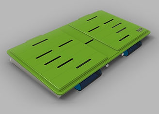
The Ophthalmic Enucleation Kit is an assembly of specially designed products that help in extraction and temporary preservation of corneas post the death of the donor.
The Need:
Over and above the under-developed countries, the Indian sub-continent has the maximum number of visionless in the world. 75% of this blindness, can be cured through transplantation. On an average, the corneal collection amounts to 0.01% of the actual need, of which only 70% is transplantable.
One of the main reasons for such a dilapidated collection rate is the loss of usable corneas, post extraction.
This loss occurs due to:
-
Inadequate transportation, sterility and access
-
Unorganised compilation of the instrument box
-
Lack of disaster management facilities.
These problem findings led to the proposed design of an Ophthalmic Enucleation Kit, 'Cornea Care', that ensures safe enucleation of the corneas and its preservation up till the nearest Eye Bank.
Various Components :
Surgical Hand Disinfectant
Bottle of water
Shoulder Mount Bag-pack:
Padded shoulder mount bag ensures safety of the corneas even during an accidental fall.
Separate dedicated compartments for
Documents
Stationary Surgical Hand Disinfectant
Water bottle
Head mount, battery operated light
A waste disposal bag
Essentials used during the Enucleation process.
Safety, Aesthetics, Comfort, Ergonomics, Dignity, Brand Identity

Uniform Chamber:
The Uniform Chamber contains few pieces of Body Apron, Face mask, and Head Mask. These are essential accessories that constitute the surgeon's uniform.
Cut outs in the chamber serve as visual clues to the content.

Organisation, Compact, Hygiene, Visual clue for missing pieces
Disposables' Unit:
This unit contains vials, saline, pack of sterile water, syringes and needles.
The unit is uncovered and placed into it's slot on the integrated instrument panel, prior to the start of the Enucleation procedure.


x
2


10
8
x
x
Organisation, Ease of access, Protection, Faster
Instant Ice Packs:
Useful for immediately securing the usability of the extracted corneas, which otherwise would not be useful for transplantation, due to delay in timely refrigeration within four to six hours, post the death of the donor.
The red McCarey Kaufman Medium (M-K.Medium) bottles, serve as sterile cages to the floating corneal button.

Disaster Management:
A set of extra instruments to serve as immediate help during unforeseen circumstances, packed into a box for easy access and organised storage.


Planar Blades
Cautery
Trephine Blades
Suture
Material
Magnifying Glass
Eye shells
Additives:
A set of instruments and aids that are specially organised for fastest access along with avoiding accidental pricking to the user.


Hand Gloves

Needle
Preservation, Storage, Longetivity
Sterile Scissors
How it works?
Eye Pads


Death of the Donor
a
Intimation to the
nearest Eyebank
b
Enucleation Kit Ready for use
Medical Practioner mounts the kit
c

Shoulder mounting helps safe transit on 2-wheelers for faster commute. It also helps in reaching to the remote areas where vehicular reach is difficult to extract corneas.


d

Documents handed to the Donor's relatives for consent along with verification and collection of Death Certificate.
e

Medical practioner orients himself/herself behind the Donor's head.
(Indian sub-continent observes a norm to place the dead body onto the floor immediately after the death)
f

Medical practioner begins
the surgery while sitting on the floor.
g

h
Corneas stored into sterile cages.

Safe storage into the
Eye bank for immediate transplant.
i
Corneal Transplantation into the Recipient.
Elements, Components and Design Features
With the use of the Cornea Care, the usability of the Corneas is preserved upto 100% till 96hours to 10 days from extraction.

Ergonomics, Safety, Organisation, Disaster Management
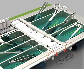
Instrument Panel:
A unique amalgamation of the sterile feild, sterile plane, sterile case and instrument box to reduce the number of sequential actions and time required to set up the environment prior to the beginning of extraction of corneas and preserve the sterility of the box.

Design Features of the Instrument Panel
Ergonomics, Safety, Sequential Organisation, Faster
Unfolding the Instrument Panel:
-
The sterile case is designed to preserve sterility of the interior environment and showcase the brand of the device/sponsering company/Eye Bank. The colours are chosen so as to negate the loud impact of the 'Red' blood post the extraction of corneal button or eyes.
-
The sterile case has strategically placed slits, which aid better autoclave sterilisation process for consecutive multiple use.
-
The ridges on the blue cover fit exactly into the slits in the green lid, to preserve the sterility, post autoclaving, till the kit is opened for surgical use.
-
The sterile case is opened by pulling the two ears towards the top edge of the blue flap.
1
2

-
The blue flap is folded below the base and the ears fit exactly into the negative space on the base of the instrument panel. The protrusion of the ears, when folded below, give an inclination of 10 degrees from the base towards the user.
-
The green cover is unveiled and folded under the base pivoting along its top edge.
-
The green cover folds under the blue flap.
-
The Disposables' Unit is inserted onto the dedicated slot in the instrument panel for easy access.
-
The two knobs on the instrument panel are rotated once to elevate all instruments at a progressively increasing angle for easy prick-free and obstruction-free access.
3

4

5

6

7

-
The sterile cage has strategically placed slits, which aid better autoclave sterilisation process for consecutive multiple use.
-
The ridges on the blue cover fit exactly into the slits in the green lid, to preserve the sterility, post autoclaving, till the kit is opened for surgical use.
-
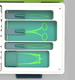
Silicone mat spread on the bottom of the 316L Stainless steel instrument panel to avoid rattling and damage to the tips. Green colour is used to dilute the effect of red blood.
-
Graphical representation of the instruments on the Silicone mat, helps the lab assistant to insert the instruments in their respective slots, while assembling the kit.
-
The cylindrical lever is pulled and rotated to lock. This lifts the instruments in a progressively increasing angle.
-
Silicone lids fold under the instrument panel to generate a forward inclination, during the surgery
Components of the Instrument Panel
Cornea Care


Half curved needle
and suture material
Slot for keeping the sterile cages, during the surgery
Enucleation Spoon
Toothed Forceps
Needle Holder
Eye Pads
Slot for keeping the Disposables' unit.
Strabismus Muscle hook
Enucleation Scissors
Eye Speculum
Dental cotton Rolls
Conjunctival Scissors
Cotton Balls

Disposables' tray placed on the instrument panel during the Enucleation process.
The sequential arrangement of the instruments with progressively increasing operational inclination.